Posts tagged bloom

High Impact Science in Antarctica

The Mertz Glacier as seen in 2007, extending 75 km out into the Southern Ocean

Antarctica was in the news this weekend when a 97 kilometer long iceberg the size of Luxembourg collided with the floating Mertz Glacier, breaking the famous glacier off at the base and generating a 2500 sq. kilometer iceberg. Each of these behemoths weigh several hundred billion tons, so the impact must have been quite a crunch!
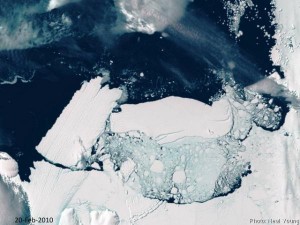
Iceberg B9B collides with Mertz Glacier Tongue

At right is an image taken February 20th, several days after the impact: the broken Mertz Glacier Tongue is on the left side of the photo, and the colliding B9B iceberg is near the center-right. The Mertz Glacier, which was sheared off at the base, was a significant barrier to westward drifting sea ice. The Mertz Glacier is on the George V coast of East Antarctica, a region is famous for its high-velocity katabatic winds: sustained wind velocities at nearby Dumont D’Urville have reached 199 m.p.h! These winds blow the pack ice out to sea, and because of the blocking geometry of the Mertz Glacier, this area generally remains ice-free all winter.
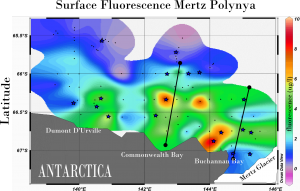
Fluorescence map of the Mertz Polynya in December 2007 (mertz Glacier is in lower right). Surface blooms are in red, and marine metagenomic samples were taken in areas marked with a star.

In the Austral summer of 2007, scientists from the J. Craig Venter Institute visited this ice-free area, or polynya, as part of the International Polar Year’s Census of Antarctic Marine Life (CAML). Because sunlight can freely penetrate the water column, polynyas are areas of enhanced productivity. Diatoms and other phytoplankton form massive springtime blooms, supporting whales, penguins, and much of the Antarctic food chain. Above is a fluorescence ‘bloom map’ of the Mertz Polynya, just west of the Mertz Glacier. Our expedition on board the Aurora Australis attempted to capture a biological snapshot of the entire region, and Jeff Hoffman and I were able to collect samples ranging from thick blooms of Phaeocystis antarctica to oligotrophic cold-water upwellings at the base of the Mertz Glacier.
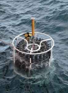
CTD Rosette being deployed at the base of the Mertz Glacier to collect a sample from 1320m deep

CTD Rosette being deployed at the base of the Mertz Glacier to collect a sample from 1320m in depth

The region around the Mertz Glacier is equally famous as one of three regions where Antarctic Bottom Water is formed (the other two are the Ross and Weddell Seas). Bottom water is created where saline water is extruded from newly formed sea ice. This cold dense water sinks from the surface and becomes distributed into all of the world’s major ocean basins. Because the sea-ice in a polynya is continuously formed and blown out to sea, there is near continual production of brine and bottom water. While in the Mertz Ploynya, Jeff and I used the ships 24-bottle CTD rosette to sample some of this bottom water, and one of the samples came from Buchanan Bay, right next to the area where the glacier split. This sample came from a depth of 1320m, and may yield insight into bacterial activities at the base of the water column. Additional deep water samples were taken in the Adelie Depression , the Mertz Bank, and the Mertz Depression, and one sample came from a depth of 3,690 m in the Southern Ocean.

Almost half of the water samples we collected have been sequenced using 454 sequencing technology and are in the process of annotation. This biological data will form an important baseline as this region undergoes rapid change: loss of the protective geometry of the Mertz Glacier will likely cause changes in the formation of the Mertz Polynya, influencing both the biology of the annual spring bloom and the dynamics of bottom water formation. Stay tuned for more updates on this exciting event and on the microbiology of the region.
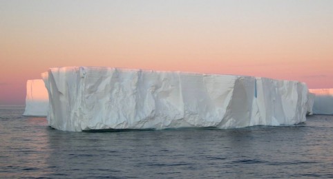
Tafelbergs floating in the morning light, Mertz Polynya, December 2007

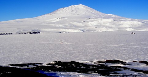
Station II, Inaccessible Island

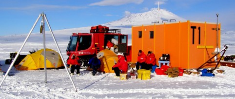
Our vehicles at Station I (near the center of the photograph)

Our camp at Station I (near the center of the sea ice field)

The second storm of our trip hit us while we were packing up Station I for a return to McMurdo. The winds began gusting over 50 miles per hour, and the visibility dropped to near zero. We had already packed up camp, but the orders came in over the radio that Condition 1 had been imposed on the sea ice route, and we were stuck there until conditions improved. Three of us slept on the floor of the research sled while Mak and I slept in the back of the Pisten Bully. The wind shook and buffeted the vehicle all night, and at times the Pisten Bully made this vibrating sound like we were just about to take off.

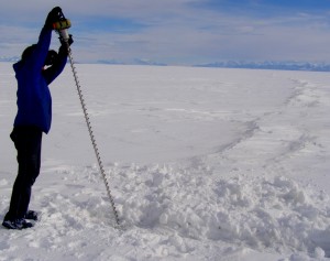
Testing ice thickness at a buried sea-ice crack

Matt drills to test the ice thickness across a crack

But by 6 PM the following day, the visibility had improved enough for us to follow the flags along the sea-ice highway and return to McMurdo. In town we picked up another crewmember: Matt Smith, the sea-ice specialist for the US Antarctic Program in McMurdo. We then drove back out to our camp at Station I and spent several hours digging our vehicles out of the snowdrifts. By noon the following day everything was ready again for redeployment, and we set out across the ice for our next station, on the north side of Inaccessible Island.

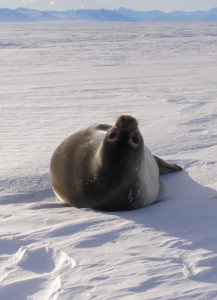
Weddell seals lounging on the ice are a warning sign for potential cracks

Weddell seals on the ice are a good indicator for nearby ice cracks

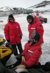
The fresh snow had buried many of the obvious ice cracks and features, so Matt and I went ahead on snowmobiles to scout the route while Jeff Hoffman and Mak Saito followed with the sleds. Cracks like the ridgeline in the above photo were relatively easy to spot, and we drilled them to make sure they were a meter thick, which is more than enough to support the weight of our vehicles. Other cracks though were less apparent, but many times those cracks were given away by the presence of seals loafing on the ice - the pup in this picture barely even moved as we rumbled by, and we saw his breathing hole in a hidden crack just a few feet away. We gave that area wide berth. After a few hours of crack testing and route finding, we made it out into McMurdo Sound proper and to our next station.

An ice core showing the diatoms growing on the bottom of the sea ice

An ice core showing the diatoms growing on the bottom of the sea ice

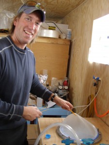
The next morning we fired up our generator and drills. I used the Kovacs core sampler to create a large enough hole so that Jeff and I could get our sampling gear down below the ice. We have been wrapping all of our sample tubing in black insulation, as the seawater will rapidly freeze on contact with icy air. This is espeically true in Antarctica, where the wind seems to blow nearly continuously, freezing engines, air hoses, compressors, you name it! I also drilled a number of ice cores so we could obtain some genetic material from the organisms living on the bottom of the ice. Drilling those cores takes a few hours- while I was doing that Jeff Hoffman worked the stainless steel Jeff Hoffman high biomass filterfilter sets and the viral concentrator. In the picture on the left you can see one of the filters for the larger phytoplankton. That particular filter captures anything in the water which is between 3 and 200 microns, which is the size of most of the diatoms and other large phytoplankton. If you have a sharp eye, or a good computer monitor, you can see a slight discoloration of the filter as compared to the edge - that discoloration is from planktonic cells which have become trapped on the filter. To obtain that amount of cells, we had to filter over 400 liters of seawater, and even then, it almost seems less that the amount that was in the ice core. This is possibly due to seasonality in the sea-ice cycle: it is still late spring here, and as summer progresses and the sea ice starts to melt, the diatoms trapped in the sea ice will be released into the water, becoming the seeds for the annual summer phytoplankton bloom in the Ross Sea. Jeff Hoffman and Andy Allen brought back samples last year from the late summer, so between the spring and summer samples we should be able to develop a wider genomic understanding of polar marine phytoplankton.

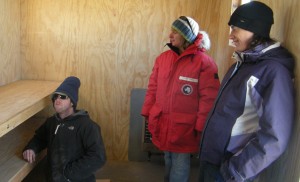
Sea-ice camp at Station II. We used the sled and the vehicles as wind barriers     Taking a break and having some hot beverages at our sea-ice camp at Station II.   We used the sled and the vehicles as a windbreak in case the weather changed.

Enjoying some hot beverages out of the wind at Station II ice camp

Ice diatoms!

Last-minute adjustments to our mobile science sled

Last-minute adjustments to our mobile science sled

Today has been a day of preparations, as tomorrow we hope to leave McMurdo Station and head out on the sea ice. Our mobile sled is almost ready for deployment: the carpenters who work for the US Antarctic Program are quite amazing, and our sled has filtration racks for separating different sizes of plankton, incubation chambers, and a mobile clean room for trace metal analyses. All of this in a 6 x 12 foot space! I’ll have more photographs in the posts to come, but it very much a sled of the 21st century.

Dawn and Abigail check out the row of Scott tents available for check-out at the Berg Field Center

We are also assembling our equipment list for camping on the ice, and the list is impressive: tents, coleman stoves, fuel, kitchen boxes, ice screws, shovels, jerry cans, carabiners, spare sleds, and a hurdy gurdy. Don’t ask me what that last thing is, I’ve been told it is a ‘fuel transfer unrelated to the musical instrument. The place to get all of these items is the Berg Field Center, which is a storehouse of mountaineering and back-country equipment available to all scientists doing Antarctic field work. For someone who likes mountaineering as much as I do, visiting the Berg Field Center is like letting loose a kid in a candy store - the place just oozes exploration.

Dawn and Mak learn about drive belts

Dawn and Mak learn about drive belts and engine parts

In the afternoon our group attended a class on snowmobiles use and maintenance. Our research sled will be pulled by a Tucker Sno-Cat, and our lighter open-air equipment sleigh will be pulled by a Pisten Bully. Both of these machines are major workhorses in and around Antarctica, but they are slow: the Pisten Bully moves along at 6 mph. So for added flexibility we decided to bring along a pair of snowmobiles. With snowmobiles, we will be able to ride ahead and scout for hazard on the ice, as well as run back to McMurdo to pick up extra equipment or drop off samples. Driving a snowmobile is similar to riding a motorcycle, although the snowmobile doesn’t have any real gears, and snowmobiles reek of partially burned fuel (dirty two-stroke engines). As part of our training we ran a mogul course, and learned how to ride on ice and on slopes, and now I understand we are all certified ‘snow machine technicians’.

Diatoms growing in the ice

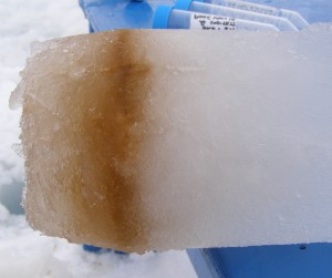
A layer of plankton growing in the sea ice

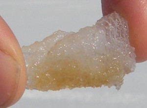
Hard to believe, but in between training sessions and lab set-up, Dawn has been able to have a look at some of the diatoms that Abigail and I brought back from our sea-ice training (see yesterday’s blog). On the right is a picture of a piece of ice from one of the holes we drilled when we were determining the thickness of the ice in cracks, and you can see that there is layer of ice discolored with pigment. These are microscopic plankton living in the ice, and for something non-mobile, living in the ice is ideal in some ways. Plankton embedded in the ice are protected from being eaten by krill and other grazing zooplankton, and ice plankton are also at the top of the water column, so they receive maximal sunlight. We transported our samples of ice back to the lab, and Dawn was able to put them under a light microscope with a camera attached and generate this picture:

Ice Diatoms: chain forming Fragilariopsis (center), ribbons of Amphiprora (squares with oval centers), and colonies of Nitzschia (long ovals growing end-to-end)

These are all diatoms, and not only could Dawn identify all of them without a guide, she could correctly spell them. Diatoms are glass shelled plankton famous for having some of the most beautiful patterned shapes in the ocean (see an SEM image here to get the idea). Diatoms play a vital role in the global carbon cycle, as the they remove large amounts of carbon dioxide from the atmosphere before dying and sinking to the bottom of the ocean. Large blooms of diatoms and other plankton may ultimately help remove much of the greenhouse gasses resulting from fossil fuel use, and is only one of many reasons why we are down here studying them.

In the bloom…almost

Cyanobacterial blooms during the summer are reoccurring phenomena in the Baltic Sea. This summer we have already encountered the two main species responsible the blooms, Aphanizomenon sp. and the toxin producing Nodularia spumigena (see previous posts), but so far not in the abundance that would qualify as a bloom. With the help from our colleagues back at JCVI in La Jolla and the Swedish meteorological institute’s satellite tracking of algae blooms (www.smhi.se) we have been following the development and waiting for the bloom to show up. However, we are approaching the end of our Baltic Sea journey and due to the relatively cold and windy weather blooms have been moderate this year. We had almost given up on the chance of encountering a bloom and due to the clouds the satellite imagery wasn’t much help when…. Finally, a bloom! Well…almost a bloom… On our way to Kalmar, just on the southern tip of the large Öland Island we spotted a lighter streak in the water and when taking a closer look we could see that the water was filled with small whitish aggregates of some sort. In the microscope we could confirm that these aggregates were indeed cyanobacteria and Nodularia spumigena seemed to be dominating the sample. Due to the whitish colour we suspect that the filaments were dying since these types of blooms are typically more yellow in color.

After sampling we continued to Kalmar, where Professor Åke Hagström was waiting for us on the dock together with his son. I was also happy to see my own brother Anders there to greet us. He and his family are in Öland where he is visiting my parents in their summer house. It was great seeing some familiar faces!

The next morning Åke came back together with his colleague Dr. Lasse Rieman for breakfast and a tour of the boat. Jeff and Åke exchanged fascinating experiences from air sampling of microbes and Åke told us about his new appointment as head of the Swedish Institute for the Marine Environment, an exciting initiative aiming to coordinate the efforts of all four Swedish marine research centres, read more about it here.

Heading north with more daylight

After spending a couple of days visiting with my family in Stockholm, I boarded a ferry boat to Blidö and rejoined the Sorcerer II crew to head north to the Bothnian Sea. Before departing, we sampled in the bay outside Dr. Norrby’s summer house. The last days of fantastic summer weather had warmed the water to about 20º C in the little bay, and we found our first specimen of Nodularia spumigena, the most conspicuous toxin producing bloom-forming cyanobacteria in the Baltic Sea. Just like Aphanizomenon this cyanobacteria have heterocysts, cells specialized for fixing nitrogen from the atmosphere.

The cyanobacterium Nodularia spumigena. Two square-formed heterocysts with thick cell-walls are visible close to the ends of the filament.

The cyanobacterium Nodularia spumigena. Two square-formed heterocysts with thick cell-walls are visible close to the ends of the filament.

We continued to sail through the night, and the crew was amazed by the light Nordic summer nights and the sunrise at 3:30 AM. The following afternoon we reached our next sampling site, one of the Helsinki Commission, or HELCOM, monitoring stations in the Baltic Sea. HELCOM has been working to protect the marine environment of the Baltic Sea for 35 years, and the monitoring stations enable researchers to identify and quantify the effects of anthropogenic discharges/activities in the Baltic Sea.

Tomorrow we continue north and meet up with Swedish scientists from Umeå Marine Research Center (UMF).

First Sampling in Plymouth Reveals Interesting Blooms — BBC Cameras capture it all!

After a couple of days in Plymouth we were ready for the first of two intense sampling days together with the Plymouth Marine Laboratory (PML). We had heard rumours about blooms of Phaeocystis, a conspicuous bloom-former in the North Sea and English Channel. When it blooms, it turns the water reddish-brown in color, and the degradation of the gelatinous colonies may result in foaming. It was another beautiful sunny morning in Plymouth when we left Sutton Harbour and headed for one of PML’s long-term coastal sampling sites, L4 and L4 east. In addition to the permanent crew, Dr. Venter, Heather Kowalski (head of PR and communications) and Dr. Chris Dupont were onboard from JCVI. They were joined by a sampling team from PML of Dr. Jack Gilbert and two students, Nicole Bale and Ben Temperton, who were going to sample for transcriptomics, i.e. analysis of RNA to look at the expression of the genome. We also invited Dr. Dawn Field and her student Paul, bioinformatics experts visiting from Oxford, to join us.

We followed PML’s research vessel Plymouth Quest out to the sampling stations. This group, headed by Denise Cummings, conducted a suite of measurements to provide us with an incredibly detailed picture of what the sampled microbial community was actually doing.

R/V Plymouth Quest

R/V Plymouth Quest

We dropped our CTD down to just over 40 m and observed two interesting changes in temperature, oxygen and pH through the water column at 12 and 28 meters respectively and decided to take a sample at 35m and a surface sample. The step-like form of the temperature profile suggested that several storms had passed through recently, mixing the water, with quiescent weather in the intermittent periods. Potentially, the drops in oxygen and pH were the result of increases in community respiration, which consumes oxygen (just like when we humans breathe). The gradual increase in chl a with depth was due to photoadaptation, that is the plankton deeper in the water produce more of the light absorbing pigments because there is less light. Fortunately, each of these hypotheses can be directly addressed by the work being done by the group aboard the Plymouth Quest. Through collaboration with PML, we will know more about these samples than we have for any sample previously.

CTD profile from L4 east.

CTD profile from L4 east.

We were delighted to find our filters full of microorganisms after filtering 200 L of seawater from the two depths. Upon opening the filter casings, we were hit with a very tangy sulfidic aroma caused by dimethyl sulphide (DMS for short). This gas, which is literally the “smell of the sea,” is the result of marine plankton degrading dimethylsulfoniopropionate (DMSP for sanity). Most phytoplankton produce lots of DMSP and the tiny animals and crustaceans that eat them are sloppy, so you essentially have a steady supply of DMSP to bacteria. Some bacteria actually want the sulphur so they metabolize the DMSP. Others degrade it to DMS, which gives you that peculiar smell. A curious side effect of DMS is that when in the atmosphere it acts as cloud condensation nuclei. In simpler terms, lots of DMS production means lots of clouds, which reflect the sun’s energy away from Earth. Therefore, in direct contrast to the carbon dioxide, DMS is a “global cooling gas.”

Just in time for the second sampling station we were met by a BBC film crew who motored up in a RIB boat. The crew from London was onboard to film the important work of the Sorcerer II Expedition and the collaboration with PML for a new science TV series to air later this year. Onboard the Sorcerer II, Dr. Venter, Chris Dupont and Jack Gilbert did an excellent job explaining our mission and the science behind it to the interested and professional BBC reporter, who also turned out to be helpful during the sampling procedure. It’s never easy with so many people onboard the boat during these intensive samplings and having a film crew, some of whom don’t have the greatest sea legs, adds to the intensity.

Dr. Venter, Jack Gilbert and Chris Dupont being interviewed by BCC reporter.

Dr. Venter, Jack Gilbert and Chris Dupont being interviewed by BCC reporter.

Earlier in the week the JCVI and PML teams had conducted some joint interviews in Plymouth while on the dock. These included a local newspaper and a BBC radio interview.

Click here to listen to one of the radio interviews.

(post by By Karolina Ininbergs and Chris Dupont)

Days of Discovery: Plymouth, Sea Urchin Cell Division and More Plankton

After a few days of fairly rough weather and winds up to 50 knots we finally spotted land and made our way to Plymouth. With our social interactions having been restricted to a pod of pilot whales and a few tankers passing through the night, we were excited to see a welcoming committee, headed by Dr. Jack Gilbert and Dave Robins from Plymouth Marine Laboratory (PML), waiting on the dock in Sutton Harbour. We were also excited to meet up with our colleague from JCVI, Dr. Chris Dupont, who flew from La Jolla to help us coordinate and run the intense sampling sessions together with our collaborators and hosts at PML. Plymouth showed us its best side with nice, sunny weather and curious and friendly spectators down in the Barbican.

Sorcerer II arriving in Plymouth.

Sorcerer II arriving in Plymouth.

Sorcerer II arriving in Plymouth.

Sorcerer II arriving in Plymouth.

On Wednesday the 20th the Sorcerer II crew was invited for a tour of the Marine Biological Association in Plymouth. The MBA is one of the oldest marine biology research institutes in the world and has been carrying out research and education for 125 years. In conjunction with PML and number of other institutions they formed the Plymouth Marine Sciences Partnership, an initiative to bring together leading marine science and technology organizations in the region. The crew was hosted by Dr. Richard Pipe, an associate fellow at the institute and head of the Plymouth Culture Collection of Marine Algae. Richard showed us around their facilities, introducing us to some of the researchers, who were happy to talk to us about their projects, ranging form cell division in sea urchin embryos to pheromones. Another highlight of the tour was the algal culture collection, consisting of some 280 strains, the oldest one isolated 100 years ago.

We also had the pleasure of visiting the National Marine Biological Library (NMBL). Linda Noble, Head of Library and Information Services, showed us some extraordinarily beautiful historical drawings of marine plankton. We were all amazed by the exquisite drawings and how well preserved the books were. Crew member Jeremy Niles put it all in historical perspective when he said, “Some of these books are older than our country!”

As our tour was coming to an end we made a final stop at the Sir Alister Hardy Foundation for Ocean Science (SAHFOS), the foundation in charge of the continuous plankton recorder survey. This marine monitoring programme has been collecting data from the North Atlantic and the North Sea on the ecology and biogeography of plankton since 1931 and is the world’s longest running marine biological survey. The continuous plankton recorders are towed by commercial ships around 17 regular shipping routes around the world every month. The data collected is used, among other things, to track global climate change, harmful algae blooms and fisheries.

Dr. Richard Pipe showing an experimental tank at the MBA to the Sorcerer II crew: Captain Charlie Howard, Jeremy Niles (Back row), Karen McNish and John Henke (front row).

Dr. Richard Pipe showing an experimental tank at the MBA to the Sorcerer II crew: Captain Charlie Howard, Jeremy Niles (Back row), Karen McNish and John Henke (front row).

The Plymouth Culture Collection of Marine Algae.

The Plymouth Culture Collection of Marine Algae.

The Plymouth Culture Collection of Marine Algae.

Captain Charlie Howard, Dr. Richard Pipe, Librarian Linda Noble and Jeremy Niles at the National Marine Biological Library.

Dr. Richard Pipe and the CPR (Continuous Plankton Recorder) at SAHFOS.

Dr. Richard Pipe and the CPR (Continuous Plankton Recorder) at SAHFOS.

We’re looking forward to sampling here in Plymouth with our PML colleagues and to the arrival of Dr. Venter. More soon.

Karolina

Back on Land

We arrive in Ft. Lauderdale and are all glad to be back on land for a few days. But we were also elated by the success of the first part of the expedition. This first journey was difficult because we had to deploy and test new equipment, to sample a diverse array of environments and oceanographic features, from large surface and subsurface blooms of photosynthetic organisms to nutrient-depleted areas of the Caribbean, and it was the first time in a year that the Sorcerer II had really been tested in open water and long distances. Data on both photosynthesis and respiration were captured, much of which will be novel and highly useful in explaining the metabolic pathways and biological participants involved in carbon and nutrient cycling in the ocean.

This week we prepare to depart for Bermuda and the Azores and will continue on to Plymouth Marine Laboratory in England. Based on the sampling success of the first leg of our journey, it is difficult to contain our enthusiasm over the microbial discoveries that lay ahead. Stay tuned as we share more scientific adventures with you.

Costa Rican Dome

In Nicaraguan waters is a regular spring upwelling event sometimes referred to as the Costa Rican dome. Winds blow across the Central American Isthmus near Lake Nicaragua and contribute to an upwelling of nutrient rich waters. These nutrients enable phytoplankton to grow, and as we approach the southern end of Nicaragua, the water takes on a greenish hue, and we note large amounts of sea turtles on the surface of the water. The turtles don’t seem to pay us much attention as we stop and take a sample. At 11 meters is a thick band of chlorophyll, and the oxygen saturation at the surface of the ocean is 117%, indicating very active photosynthesis (and production of oxygen). As before, we take samples from the oxygen minimum layer and the chlorophyll max. From what I understand, this bloom has only been detected by satellite, and we are the first research group to take genomic samples from this important phytoplankton bloom.

Sampling Blooms in Cabo Corrientes

Just south of Puerto Vallarta is Cabo Corrientes, and our satellite data indicate a large bloom extending 25 miles off the coast. As we enter the bloom the water turns an intense green, and there are numerous fish feeding in the area. Sampling conditions are ideal: bright sunshine, light winds, moderate swell. We deploy a large plankton net which rapidly fills with algae and zooplankton. Karen McNish looks at the larger diatoms and zooplankton under the scope while the rest of the crew prepares our instrumentation for deployment.

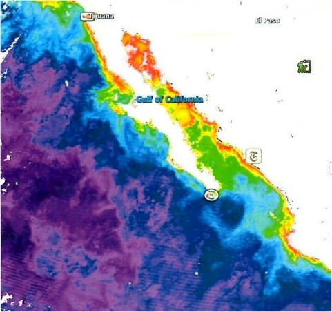
Satellite image of phytoplankton blooms along the Mexican coastline, March 2009.  The Ilsa Cedros bloom is halfway down the Baja peninsula on the west side, the Cabo Corrientes bloom is the red area in the lower right corner of the image.

Satellite image of phytoplankton blooms along the Mexican coastline, March 2009. The Ilsa Cedros bloom is halfway down the Baja peninsula on the west side, the Cabo Corrientes bloom is the red area in the lower right corner of the image.

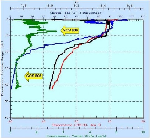
The CTD profile of the water column at Cabo Corrientes showing a surface phytoplankton bloom.

The CTD profile of the water column at Cabo Corrientes showing a surface phytoplankton bloom.

From the aft cockpit we deploy a CTD equipped with a sampling hose. A standard CTD measures conductivity, temperature and depth: our unit also contained a pH probe and a fluorometer for measuring chlorophyll concentration. As we lower the CTD through the water column, we generate a profile of the ocean at Cabo Corrientes down to 40 meters in depth. At left you can see the CTD plot: depth is plotted on the y-axis as a change in pressure, and pH (black) and temperature (red) are plotted on the top two x-axes, with oxygen (blue) and fluorescence (green) plotted on the bottom two x-axes. In this case, the peak fluorescence (green trace) is at 8 meters in depth, and after that, the concentration of oxygen (blue trace) falls from 90% saturated to 5% saturated. The peak fluorescence indicates the location of the chlorophyll max (or Chlmax), where most of the photosynthetic plankton are located, and the oxygen minimum (or O2 min) indicates an area of intense respiration immediately under the Chlmax. Both of these areas contain a wealth of undescribed microorganisms, and understanding the relationship between photosynthesis and respiration in the ocean is one of the keys to understanding the global carbon cycle. We took samples at 8 meters and at 35 meters before continuing our southward trip.